You’ve seen the posters. Usually, it’s a bright red-and-blue sketch hanging in a doctor's office or a middle school biology classroom. It looks clean. It looks organized. But honestly, if you actually saw a real heart diagram in the body, it’s a chaotic, crowded mess of plumbing that looks nothing like those primary-colored icons on your screen.
The heart isn't even where most people think it is. You’re told it’s on the left. It isn't. It’s mostly central, tucked behind the breastbone, just tilted slightly so the bottom point—the apex—pokes toward your left ribs. If you’re looking for a perfect "Valentine" shape, you're going to be disappointed. The real thing is a muscular, lopsided pump about the size of your clenched fist, and it’s arguably the most hardworking piece of meat in the known universe.
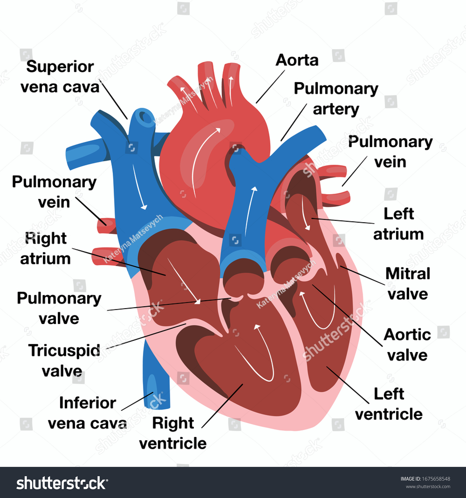
Mapping the Real Heart Diagram in the Body
When we talk about a heart diagram in the body, we’re really talking about a roadmap of pressure. The heart is essentially two pumps taped together. The right side is the "low pressure" side. It takes the "used" blood—the stuff that's already dropped off its oxygen to your toes and brain—and shoves it into the lungs. The left side is the powerhouse. It’s thick. It’s meaty. It has to be, because it’s responsible for blasting blood all the way from the top of your skull to the tips of your toenails.
Most diagrams fail to show the sheer scale of the Great Vessels. The aorta isn't just a little tube; it’s a massive, candy-cane-shaped highway that arches over the top of the heart. If that thing springs a leak, it's game over in minutes. Then you have the Superior and Inferior Vena Cava. These are the "return chutes." They dump deoxygenated blood back into the Right Atrium.
People get confused about the chambers. Think of them as waiting rooms and launchpads. The atria are the waiting rooms. They’re thin-walled because they don't have to do much heavy lifting. The ventricles? Those are the launchpads. The Left Ventricle is the MVP here. In a true anatomical heart diagram in the body, the wall of the left ventricle is nearly three times thicker than the right. It’s built for war.
The Electrical Grid You Never See
If you just look at the muscle, you’re missing the "software" that runs the hardware. Every heart diagram in the body should technically include the electrical conduction system, but most leave it out because it’s hard to draw.
It starts with the SA node. This is your natural pacemaker. It’s a tiny clump of cells in the right atrium that decides how fast you're going to live today. It sends a spark—literally an electrical impulse—that travels through the heart walls. This spark tells the atria to squeeze first, then hits the AV node (the "gatekeeper"), pauses for a fraction of a second to let the blood actually move, and then triggers the ventricles.
Without that tiny pause at the AV node, your heart would just quiver. It wouldn't pump. This is what happens during Ventricular Fibrillation (V-fib). The rhythm goes haywire, the "map" breaks, and the pump stops. This is why AEDs exist—to "reset" the electrical diagram and get the SA node back in charge.
The Valves: The Unsung Security Guards
You have four main valves. They are the swinging doors of the heart.
- Tricuspid and Mitral: These sit between the atria and ventricles.
- Pulmonary and Aortic: These sit at the exits.
Ever heard a heartbeat? Lubb-dupp. That isn't the sound of muscle squeezing. It’s the sound of these doors slamming shut. The "Lubb" is the Mitral and Tricuspid valves closing so blood doesn't backflow into the "waiting rooms." The "Dupp" is the Aortic and Pulmonary valves snapping shut after the blood has been ejected. If these doors leak (regurgitation) or get stiff (stenosis), the whole heart diagram in the body becomes inefficient. Your heart has to work twice as hard to move the same amount of blood. It’s like trying to bail out a boat with a leaky bucket.
Why the "Blue Blood" Myth Persists
We need to address the colors. In almost every heart diagram in the body, the veins are bright blue. This has led generations of kids to believe their blood is blue until it hits the air.
It’s not.
Blood is always red. When it’s full of oxygen, it’s a bright, cherry red. When it’s "empty" or deoxygenated, it’s a deep, dark maroon. It only looks blue through your skin because of the way light interacts with your tissue and the vessel walls. Doctors use blue in diagrams simply because it’s a convenient way to show the difference between "fresh" and "used" blood. If they used two shades of red, you’d never be able to tell what’s going on in a complex drawing.
The Coronary Arteries: The Heart's Own Supply Line
Here is the irony: the heart is full of blood, but it can’t use any of it. The blood passing through the chambers doesn't nourish the heart muscle itself. The heart needs its own private plumbing system.
These are the Coronary Arteries. They wrap around the outside of the heart like a crown (hence the name "coronary"). In a detailed heart diagram in the body, you’ll see the Left Main Coronary Artery branching off into the LAD (Left Anterior Descending). Doctors call the LAD the "Widowmaker." Why? Because it supplies blood to the front of the left ventricle. If that specific pipe gets blocked, the most important part of the pump dies.
Realizing this changes how you look at a diagram. It’s not just a pump; it’s a pump that is also its own customer. If the pump doesn't feed itself first, it stops pumping for everyone else.
What Most People Get Wrong About Placement
You’ve probably seen someone clutch their far left chest during a movie heart attack. In reality, cardiac pain usually presents as a crushing weight right in the center of the chest, under the sternum.
Because the heart diagram in the body shows it nestled between the lungs, the symptoms of heart trouble often mimic other things. It can feel like bad indigestion. It can feel like a pulled muscle in your jaw or your left arm. This is called "referred pain." Your brain gets its wires crossed because the nerves from the heart and the nerves from your arm enter the spinal cord at the same level.
Evolution and Variations
Not every heart diagram in the body looks the same. Some people are born with "Dextrocardia," where the heart is flipped and points to the right. Others have "holes" in the heart (Atrial Septal Defects), where the wall between the two sides didn't close properly after birth.
In a fetus, the "diagram" is actually different. There’s a shortcut called the ductus arteriosus because the baby isn't using its lungs yet. The moment that first breath is taken, the pressure shifts, the shortcut closes, and the "adult" diagram takes over. It’s a literal mechanical shift that happens in seconds.
Actionable Insights for Heart Health
Understanding the heart diagram in the body isn't just for passing a biology test. It’s about knowing how to maintain the machinery.
- Watch the "pipes": High blood pressure (hypertension) is like turning the garden hose on too high. Over time, it scars the delicate linings of the arteries shown in your diagram, making it easier for "gunk" (plaque) to stick.
- Check the "battery": If you feel skipped beats or a racing heart while sitting still, your electrical grid might be misfiring. It’s worth an EKG to see if the SA node is behaving.
- The "pump" needs exercise: The heart is a muscle. If you don't make it work, the walls of the ventricles get flabby. Cardio isn't just about burning calories; it’s about "pressure-testing" the system to keep the muscle walls elastic and strong.
- Listen to the "valves": A heart murmur is often just the sound of turbulent blood flow through a valve. Most are "innocent," but some indicate a door that isn't closing right.
The heart is a masterpiece of engineering, but it’s a physical object subject to the laws of physics. It has a limited number of beats—roughly 2.5 billion in an average lifetime. Mapping out your own heart diagram in the body through regular checkups, blood pressure monitoring, and understanding your family history is the only way to ensure you get every last one of those beats. Don't wait for a "clutch the chest" moment to start paying attention to the plumbing.