You’re finally here. You’ve probably spent the last few weeks staring at a pregnancy test stick or refreshing a period-tracking app, but the 10-week mark is where things start to feel, well, real. It’s a massive milestone. By the time you see an image of fetus at 10 weeks, you aren’t just looking at a "clump of cells" anymore. You’re looking at a tiny human who has officially graduated from the "embryo" stage to become a "fetus."
It’s wild. Honestly, the change that happens between week 8 and week 10 is some of the fastest development in the entire human experience. If you enjoyed this post, you should read: this related article.
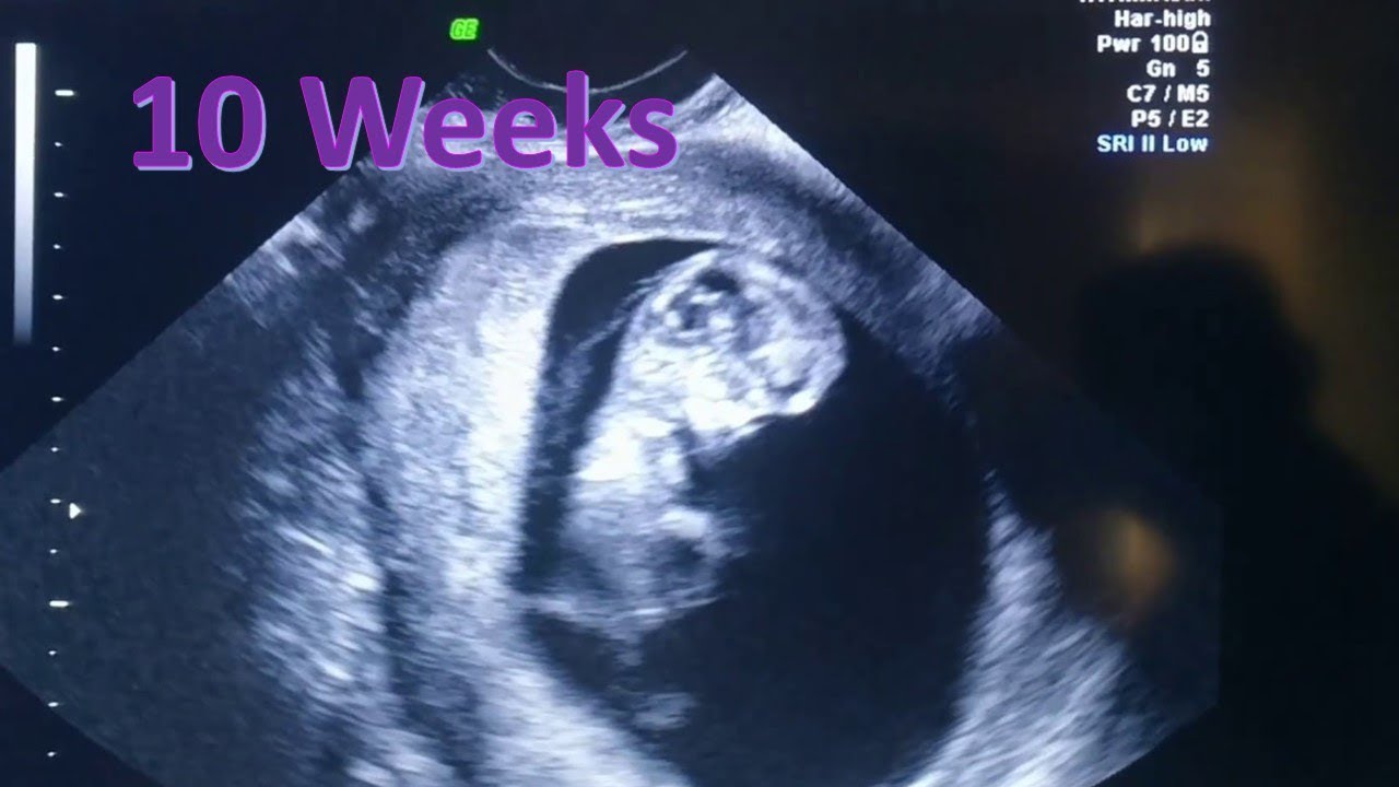
If you’re heading into an OB-GYN office or a boutique imaging center, you might be expecting a clear, portrait-style photo. Reality check: it’s usually a bit more abstract than that. Depending on the equipment and the position of your uterus, that image of fetus at 10 weeks might look like a gummy bear doing jumping jacks or a very blurry space alien. But to a trained sonographer, that grainy gray shape is a roadmap of incredible biological progress.
What You’re Actually Seeing in a 10-Week Ultrasound
Most people expect to see a face. You won't. Not really. At ten weeks, the head is still disproportionately huge—it’s basically half the size of the entire body. This is because the brain is growing at an astronomical rate, producing about 250,000 new neurons every single minute. For another angle on this story, see the latest update from Medical News Today.
When you look at the image of fetus at 10 weeks, look for the "C" shape. The spine is starting to straighten out, but there’s still a distinct curve. You might see tiny paddles. Those are the hands and feet. Last week, they were webbed. Now? The webbing is disappearing, and individual fingers and toes are forming. They’re still minuscule, but they’re there.
The tail is gone. That’s a big one. The "embryonic tail" at the bottom of the spinal cord has been reabsorbed, making the fetus look much more like a miniature person.
Transvaginal vs. Abdominal Imaging
Look, let’s be real about the "how" of getting this image. At 10 weeks, your uterus is roughly the size of a large orange. It’s still tucked down behind your pubic bone. Because of this, many doctors still prefer a transvaginal ultrasound to get the clearest image of fetus at 10 weeks.
It’s not the most glamorous experience.
But it gets the camera much closer to the action. If they try an abdominal ultrasound (the one where they rub cold gel on your belly), and you have a tilted uterus or a bit of extra cushion, the image might be super blurry. Don't panic if they switch methods halfway through. They just want a better "window" to see the heart flickering.
The "Gummy Bear" Phase and Subtle Movements
If you get a high-resolution 2D image, the fetus often resembles a gummy bear. You’ll see a distinct head, a torso, and little nubs for limbs. But the coolest part of a 10-week scan isn't the still photo—it’s the video.
Even though you can’t feel a thing yet (and you won't for several more weeks), the fetus is moving. A lot. They’re jerky, spasmodic movements. They might bounce off the wall of the uterine sac or wiggle those new limbs. It’s involuntary, sure, but seeing that motion on the screen is usually the moment the "holy crap, I’m pregnant" feeling finally sinks in for most parents.
Behind the Pixels: Internal Development at Week 10
While the image of fetus at 10 weeks shows the outside, what’s happening inside is arguably more impressive. According to the Mayo Clinic and ACOG (American College of Obstetricians and Gynecologists), this is the week where the vital organs—liver, kidneys, intestines, and lungs—are starting to function. They aren't "finished," but they’re online.
- The Heart: It’s fully formed and beating at a frantic pace, usually between 140 and 170 beats per minute. On the ultrasound screen, this looks like a rapid, rhythmic flickering.
- The Gut: This is a weird fact most people don't know: the intestines actually develop inside the umbilical cord for a bit because there isn't enough room in the tiny abdomen. Around week 10, they start migrating back into the belly.
- The Jaw: Tiny tooth buds are already forming under the gums.
Basically, the "blueprint" phase is over. The "construction" phase is in full swing.
Why the Nuchal Translucency Scan Matters Now
You might hear your doctor mention the NT scan. This is often done between weeks 11 and 13, but some practitioners start looking at the foundations of it at 10 weeks. They are measuring the clear (translucent) space at the back of the baby’s neck.
Increased fluid in that area can sometimes be an early marker for chromosomal conditions like Down Syndrome. It’s not a diagnosis—just a screening tool. If you see the technician focused intently on the back of the neck during your image of fetus at 10 weeks session, that’s what they’re checking. It's standard procedure, but it can feel tense if you don't know why they're zooming in so much.
Misconceptions About 10-Week Images
People get a lot of things wrong about this stage. Social media is full of highly processed 3D/4D images that make it look like you can see the baby's eyelashes. You can't.
"Can I tell the gender yet?"
Technically? Yes, the DNA knows. Visually? No. On a standard image of fetus at 10 weeks, the external genitalia are still developing and look nearly identical in both boys and girls—a little nub called the genital tubercle. If you want to know the sex this early, you’re looking at a NIPT (Non-Invasive Prenatal Testing) blood test, not an ultrasound image. Don't let a "nub theory" expert on a forum convince you they know for sure based on a 10-week grainy photo.
"It looks like a fish/alien."
That’s totally normal. The eyes are currently on the sides of the head and are fused shut. The ears are very low. It takes time for everything to migrate to the "human" positions we recognize.
What if the image doesn't look like what I expected?
Sometimes the scan is disappointing. Maybe the baby is sleeping and tucked into a corner, or the angle is weird. An image of fetus at 10 weeks is a snapshot in time, not a professional headshot. Factors like the amount of amniotic fluid, the position of the placenta, and even how much water you drank before the appointment can change the clarity.
Doctors are primarily looking for:
- Crown-Rump Length (CRL): Measuring from the top of the head to the bottom of the torso to confirm your due date.
- Heartbeat: Confirming a strong, steady rhythm.
- Gestational Sac: Ensuring everything is placed correctly within the uterus.
Practical Steps for Your 10-Week Appointment
If you have your scan coming up, don't just show up and hope for the best.
Hydrate, but don't overdo it. A semi-full bladder can sometimes help push the uterus into a better position for an abdominal scan, but if it’s too full, you’ll be miserable while they press the transducer down. Follow your clinic's specific instructions.
Ask for the "Goldilocks" print. Technicians usually give you a few thermal prints. Ask if they can get a shot of the profile (the side view). This usually provides the clearest "baby-like" silhouette for your fridge or announcement.
Bring a partner or record it. If your clinic allows it, have someone record the screen when the heartbeat is playing. The sound of a 10-week heartbeat is something you’ll want to hear again when you’re feeling anxious later on.
Understand the limitations. If you’re at a "boutique" ultrasound place, remember they aren't doctors. They are for entertainment. If they see something "off" on the image of fetus at 10 weeks, they aren't legally allowed to diagnose it. Always prioritize your medical scans over the "pretty" ones.
Moving Forward After the Scan
Once you’ve seen the image and confirmed a heartbeat, the risk of miscarriage drops significantly—often to less than 2-3%. That’s a huge relief.
You’ve officially moved out of the highest-risk period of the first trimester. Take that image of fetus at 10 weeks, put it somewhere safe, and start thinking about the next steps. You'll likely be offered genetic screening (NIPT) around now, which uses your blood to look at the baby’s DNA. Between the clear ultrasound and a "low risk" NIPT result, many parents finally feel "safe" to start sharing the news with the world.
The next big visual milestone is the 20-week anatomy scan. It feels like a lifetime away, but the jump in detail between 10 weeks and 20 weeks is even more staggering than the one you just witnessed. For now, enjoy the gummy bear. It’s the busiest your "orange-sized" uterus will ever be.
Your 10-Week Checklist:
- Schedule your Nuchal Translucency (NT) scan if you haven't yet; the window closes after week 13.
- Ask your provider about NIPT bloodwork if you want to know the sex and chromosomal health earlier than the 20-week scan.
- Start a "bump" photo progression. The 10-week mark is a great baseline because the bloating might finally be turning into a tiny, firm bump.
- Check your prenatal vitamin for DHA and Folic Acid—this is the peak of brain and spinal development.
The transition from embryo to fetus is a massive biological hurdle. Seeing that first image of fetus at 10 weeks is the reward for surviving the morning sickness and exhaustion of the early weeks. It’s proof of the work your body is doing behind the scenes. Keep the photos, stay hydrated, and breathe—you're nearly through the first trimester.